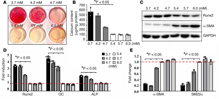
Figure 3. Lower physiological concentrations of potassium induced VSMC osteogenic differentiation and calcification.
(A) Effects of potassium levels on calcification of vascular smooth muscle cells (VSMCs), determined by Alizarin red staining. VSMCs were cultured in osteogenic media with increased concentrations of potassium, 3.7 to 6.0 mM, for 3 weeks. Representative images of stained dishes from 4 independent experiments are shown. (B) Total calcium content in VSMCs, determined by Arsenazo III. VSMCs were cultured in osteogenic media with increased concentrations of potassium, 3.7 to 6.0 mM, for 3 weeks. Results shown are normalized by total protein amount. Bar values are means ± SD (n = 3, *P < 0.05 compared with potassium at 5.4 mM). (C) Effects of potassium levels on the expression of osteogenic and smooth muscle cell markers. VSMCs were exposed to 3.7 to 6.0 mM of potassium for 3 weeks. Representative images of Western blot analysis of runt-related transcription factor 2 (Runx2) and α-smooth muscle actin (α-SMA) proteins in VSMCs exposed to different concentrations of potassium from 3 independent experiments are shown. (D and E) Real-time PCR analysis of (D) osteogenic markers, Runx2, osteocalcin (OC), and alkaline phosphatase (ALP) and (E) smooth muscle cell markers, α-SMA and smooth muscle protein 22 α (SM22α). VSMCs were exposed to 3.7 to 6.0 mM potassium for 10 days. Results from 3 independent experiments performed in duplicate are shown. Bar values are means ± SD (*P < 0.05 compared with potassium at 5.4 mM). Statistical analysis was performed by 1-way ANOVA followed by a Student-Newman-Keuls test.

