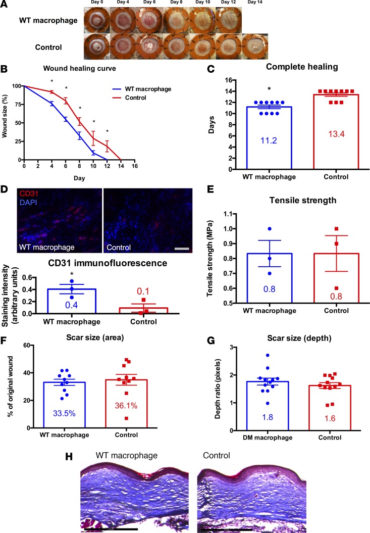
Figure 1. Macrophage transplantation improves cutaneous wound repair in a humanized model of excisional wound healing in wild-type mice.
(A) Gross photographs of wounds (n = 10 per group) during healing with macrophage-seeded hydrogel (top row) or with hydrogel control (bottom row). (B) Wound healing curve showing wound size as a percentage of original wound versus time in days (*P < 0.01; 2-tailed unpaired Student’s t test; n = 10 per group). (C) Bar graph showing the difference in time to complete healing between macrophage-treated and control wounds in wild-type mice (*P < 0.001; 2-tailed unpaired Student’s t test; n = 10 per group). (D) CD31 immunofluorescence of fully healed wounds treated with macrophage-seeded hydrogel (top left) versus hydrogel control (top right) and bar graph with immunofluorescence quantification (bottom; *P < 0.05; 2-tailed unpaired Student’s t test; n = 3 per group). Scale bar: 100 μm. (E) Bar graph of tensile strength in fully healed wounds treated with macrophage-seeded hydrogel versus hydrogel control (P > 0.05; 2-tailed unpaired Student’s t test; n = 3 per group). (F) Bar graph of scar size as a percentage of original wound in fully healed wounds treated with macrophage-seeded hydrogel versus hydrogel control (P > 0.05; 2-tailed unpaired Student’s t test; n = 10 per group). (G) Bar graph of scar size as depth ratio to normal unwounded skin, as seen in histology of fully healed wounds treated with macrophage-seeded hydrogel versus hydrogel control (P > 0.05; 2-tailed unpaired Student’s t test; n = 10 per group). (H) Masson’s trichrome stain of fully healed wounds treated with macrophage-seeded hydrogel (left) versus hydrogel control (right). Scale bar: 500 μm.

