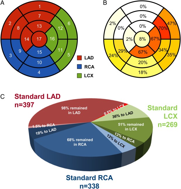
Figure 2.
(A) Standardized myocardial segmentation model used in this study with number codes for each segment (see Table 3).14 (B) Reassignment rates by hybrid imaging for the 1004 pathological segments (the intensity of colours in each segment indicates the frequency of reassignment of that segment when pathological). (C) Pie chart indicating proportion of reassignment and reassignment fate for pathological segments in each standard coronary territory. Shades of red indicate standard LAD, of green standard LCX, and of blue standard RCA territories. Standard LCX segments were most often reassigned to LAD (36%), while standard RCA segments were equally distributed between LAD and LCX.

