Figure 3.
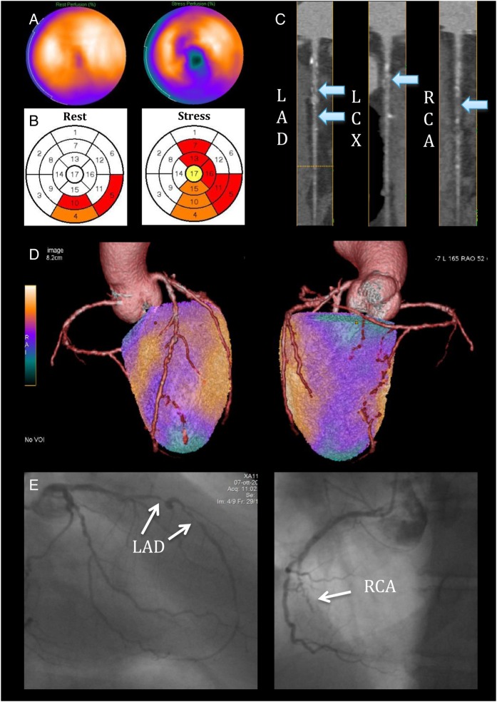
A 55-year-old gentleman with atypical chest pain. (A) SPECT shows a reversible perfusion defect inferiorly with lateral extension, and in addition, there is a separate reversible perfusion defect involving the apical region and the mid-ventricular anteroseptal wall. (B) The perfusion polar maps show the SPECT core-lab interpretation (white = normal, yellow = mildly reduced, orange = moderately reduced, and red = severely reduced radiotracer uptake) with pathological segments assigned to all three coronary territories. (C) CTCA reveals two 70–90% mid LAD stenoses, a 50% proximal LCX stenosis, and a probable occlusion of the mid RCA (arrows). (D) On hybrid imaging, the entire inferolateral perfusion defect is reassigned to the RCA, effectively changing the diagnosis from three-vessel to two-vessel disease. (E) Imaging findings were confirmed on QCA showing two high-grade lesions in the mid LAD, diffuse non-significant disease in the LCX, and a chronic total occlusion of the mid RCA.

