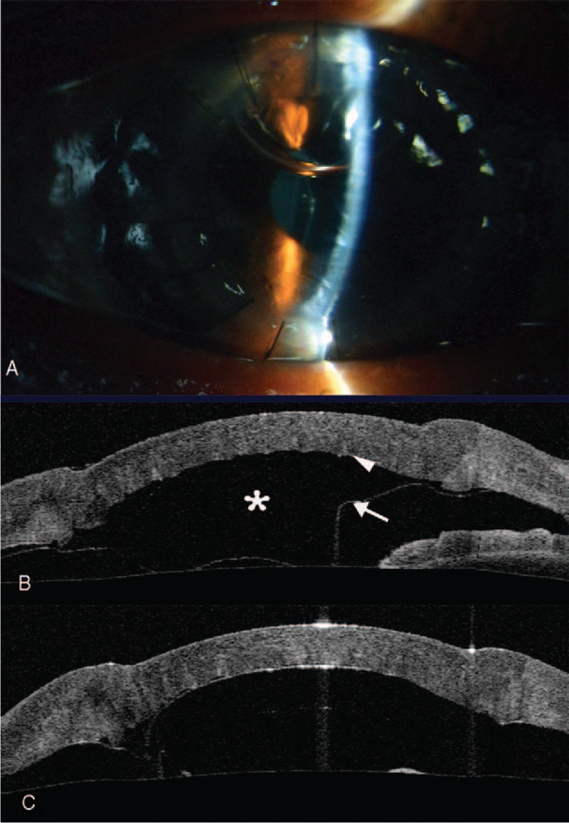
Figure 2.

One-day postoperative appearance of the right eye showing DM detachment and a double anterior chamber. (A) Slit-lamp image and (B) AS-OCT image. (C) AS-OCT immediately after the air injection. The image showed that the resolution of DM detachment resolved except for a circumscribed detachment. Asterisk: detachment pool. Arrowhead: entrance pump. Arrow: exit pump. AS-OCT = anterior segment optical coherence tomography, DM = Descemet membrane.
