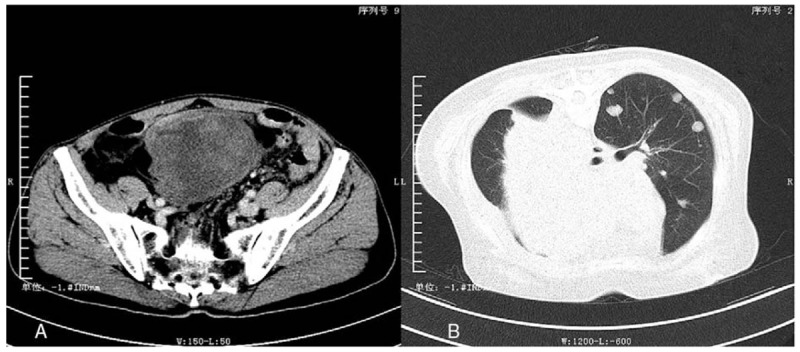
Figure 1.

The enhanced computed tomography (CT) of primary pleomorphic liposarcoma (PLS) located in lower abdomen (A) and the CT scan of metastatic lesions (B). A, The picture is the enhanced CT of case no. 4. There was a large soft-tissue shadow in lower abdomen, with heterogeneous microenhancement and clear boundary. B, The picture is the CT scan of the case no. 5 before her last surgery. There were couples of unequal-sized metastatic tumors in the left side of lung and a giant metastatic tumor in the mediastinum.
