
Figure 1.
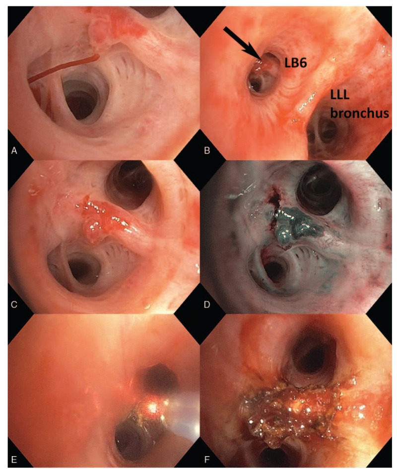
Active bleeding into saline-filled bronchus (panel A). Lesion location on proximate secondary carina of the superior segment of the left lower lobe (panel B). White light (panel C) and narrow-band imaging (panel D) of the lesion after hemostasis was achieved. Destruction of the lesion with Nd:YAP laser, starting with the afferent region at 3 o’clock (panel E). Final result (panel F).