
Figure 2.
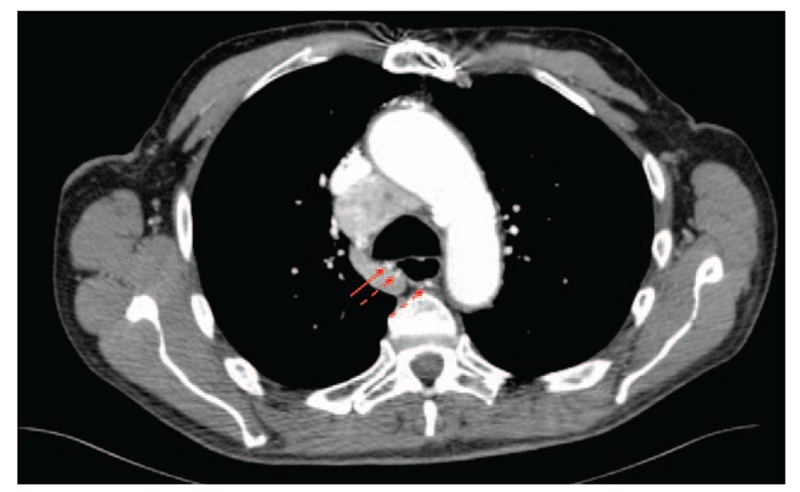
Axial cut of an intravenous contrast-enhanced computed tomography scan of the chest, demonstrating a tortuous hypertrophied bronchial artery (3 arrows) supplying a large subcarinal metastasis not visualized in this image. The vessel approximates the posterior membrane of the proximal right mainstem bronchus (solid arrow) at the location of the bleeding lesion identified at bronchoscopy.