
Fig. 3. Glioblastoma EVs contain PD-L1 that directly interacts with PD1, and the interaction is displaced by anti-PD1 treatment.
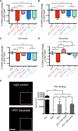
(A and B) PD1 blockade prevents the inhibition of PD-L1high GSC EVs on PBMCs. Percent change in CD69 expression for CD4+ (A) and CD8+ (B) T cells. PD1 blocking antibody (10 μg/ml) or isotype control (10 μg/ml) was added at day 0 (n = 7 PBMC donors, means ± SD). (C and D) PD1 blockade furthermore prevents the inhibition of PD-L1high GSC EVs on CD3+ isolated cells. CD3+CD4+ (C) and CD3+CD8+ (D) cells (n = 3) after treatment. (E) PD-L1–carrying, palmtdT-labeled PD-L1high GSC EVs can bind to wells coated with recombinant PD1, whereas PD1 antibody blockade inhibits EV binding. Representative confocal images are shown on the left, whereas quantification is provided on the right. Spots per field of view (FOV) on the y axis represent palmtdT-positive dots. Scale bar, 50 μm; ×500 magnification inserts; quadruplicates as means ± SD. One-way ANOVA, with post hoc Bonferroni’s correction, was used to differentiate multiple groups (****P < 0.0001, ***P < 0.001, **P < 0.01, and *P < 0.05).