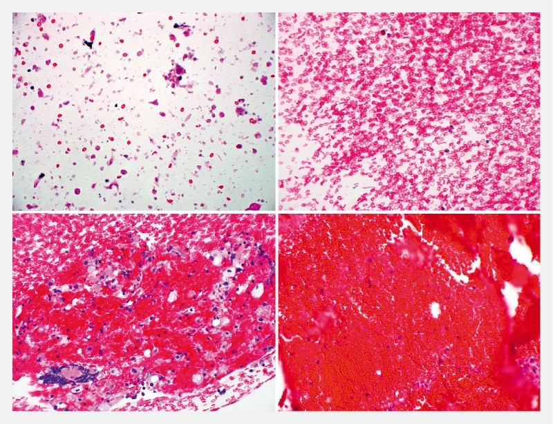
Fig. 1.

Grading scale of blood of cell block specimen. Photomicrographs show grading of the degree of blood present in a specimen. 0 (A): nearly absent of RBC; 1 + (B): monolayer of RBC, no cluster formation; 2 + (C): aggregates of RBC, < 1 per high power field (HPF) (x400); 3 + (D): aggregates of RBC present, > 1 per HPF (× 400)
