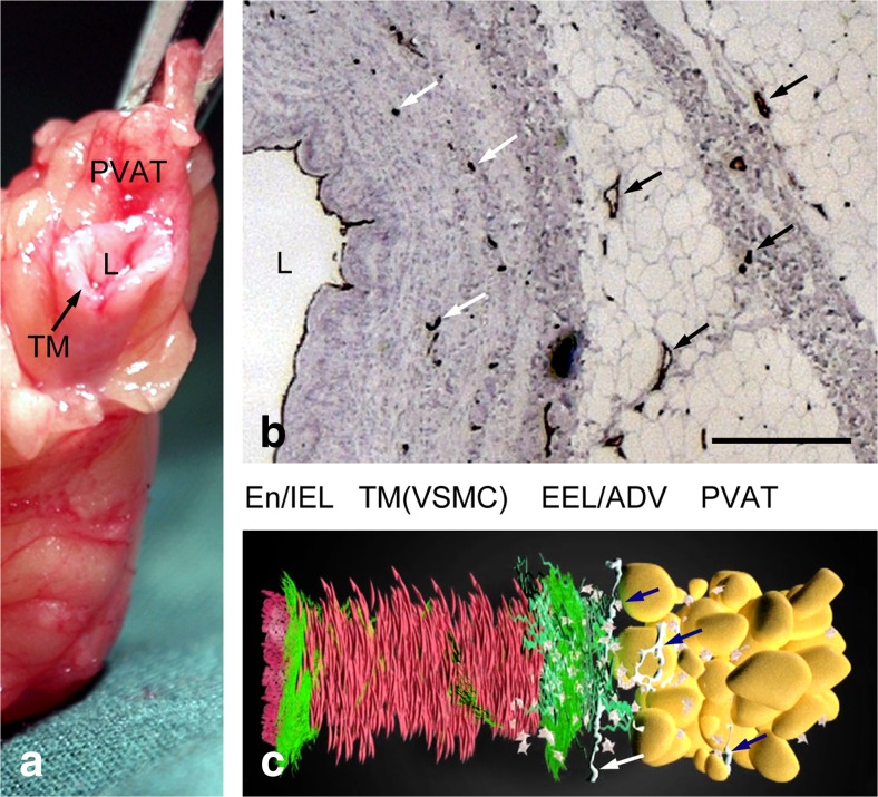
Fig. 1.
Structure of vessel wall of human saphenous vein. a A length of SV harvested in preparation for use as a graft in a patient undergoing CABG; L-lumen, TM-tunica media, PVAT-perivascular adipose tissue. b Light microscopy of a part of a transverse section of a human SV used for CABG immunolabelled for endothelial cells (CD31 antibody: brown staining) lining the lumen (L) and vasa vasorum (arrows) of the media, adventitia and PVAT (from MR Dashwood unpublished). Bar: 250 μm. c Animated reconstruction of a human SV used for CABG showing the layers of the wall and nerves (arrows); End/IEL-endothelium/internal elastic lamina, TM(VSMCs)- vascular smooth muscle cells of the tunica media; EEL/ADV-external elastic lamina/adventitia. (We gratefully acknowledge Dr. Craig Daly and Ms. Anna Mikelsone at www.cardiovascular.org for permission to use this image; see Mikelsone (2014))

