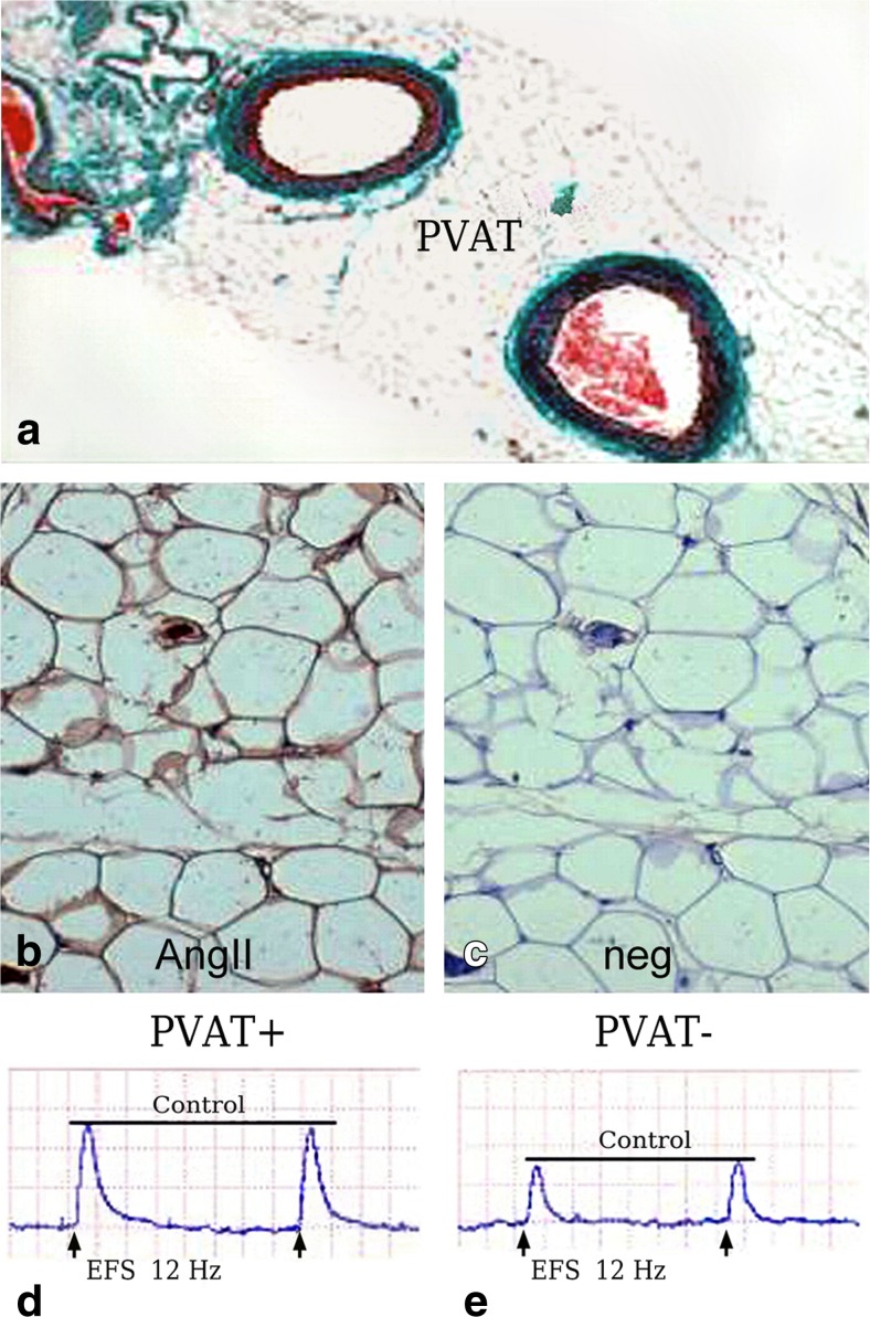
Fig. 2.
Location of angiotensin II (AngII) in PVAT and its effect on nerve stimulation. a Sections of rat mesenteric arteries surrounded by PVAT. b Presence of AngII by immunohistochemistry (brown stain); c negative control (neg) for AngII staining. d-e Representative constrictor responses to electrical field stimulation (EFS) in arteries with PVAT intact (PVAT+) and PVAT removed (PVAT-); there is attenuation of the constrictor effect when PVAT is removed. (a-e Modified from Lu et al., Eur J Pharmacol 2010, 634(1–3):107–112 [Elsevier] with permission, which we gratefully acknowledge)

