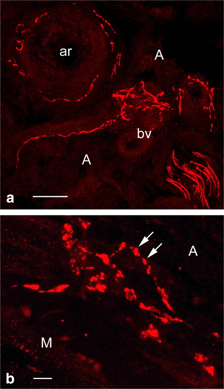
Fig. 4.
Confocal microscopy of 30 μm frozen cross-sections of non-stripped and non-distended (control) human SV graft preparations for CABG (~ 30 min to harvesting) with preserved paravascular connective tissue including white adipose tissue displaying immunolabelling for tyrosine hydroxylase (TH) in sympathetic nerve fibres (red). a Note sympathetic nerve fibres in the outer adventitia (A) and in the vicinity of vasa blood vessels (bv) including an arteriole (ar). Bar: 50 μm. b At higher magnification note varicosities (arrows) of sympathetic nerve fibres at the adventitia (A) – media (M) border. Bar: 10 μm. [Note that a rabbit polyclonal anti-TH antibody (TZ 1010, Affinity, Exeter, UK) was used at 1:300 dilution. Goat anti-rabbit Alexa 568 (Molecular Probes, Oregon, USA) was used at 1:600 as a second layer. Confocal laser microscope: BioRad Radiance 2000]. (We gratefully acknowledge that this figure was modified from Loesch and Dashwood, Phlebology Digest [Excerpta Medica Elsevier BV, Amsterdam] 2009b, 22(2):22–24)

