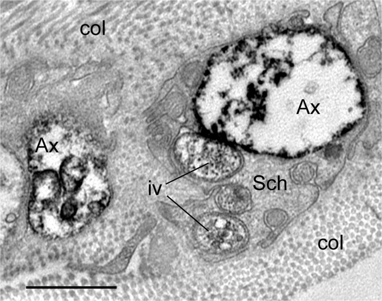
Fig. 5.
Transmission electron microscopy of perivascular nerve fibres of human SV ~ 30 min to harvesting (vein partially stripped off pedicle) showing immunolabelling for tyrosine hydroxylase (black stain) in sympathetic nerves. Note that the axon varicosities (Ax) of sympathetic nerves are damaged displaying partially “empty” axoplasm; immune-positive intervaricosities (iv) and immune-negative Schwann cell (Sch) are rather well preserved; col.-collagen. Similarly damaged axon varicosities can also be observed in non-stripped and non-dilated SV during harvesting; the proportion of axons affected in each method of harvesting is unknown. Bar: 1 μm. [Note that a TH rabbit polyclonal antibody (TZ1010, Exeter, UK) was used in the pre-embedding ExtrAvidin immunohistochemical method on paraformaldehyde-glutaraldehyde-fixed 50 μm cryostat sections, which then were embedded in Araldite; ultrathin 80 nm sections were examined with a Philips CM120 transmission electron microscope]. (We gratefully acknowledge that this figure was modified from Loesch and Dashwood, Phlebology Digest [Excerpta Medica Elsevier BV, Amsterdam] 2009b, 22(2):22–24)

