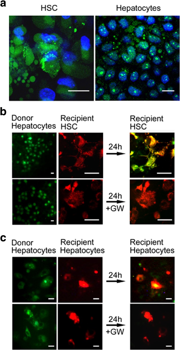
Fig. 5.
ExoHep mediate transfer of RNA into HSC or hepatocytes. a Uptake into recipient HSC or AML12 cells of SYTO RNASelect™ (green) after 24-h incubation of the cells with SYTO RNASelect™-loaded ExoHep. Blue is DAPI, green is RNA. Control cells not incubated with exosomes did not stain green (data not shown). b AML12 exosome donor cells in which RNA was stained with SYTO RNASelect™ (green) were co-cultured in 2-well μ-dish devices with PKH26-stained mouse HSC (red) for 24 h. The green/yellow color (far right, upper panel) demonstrates uptake into recipient HSC of AML12 cell-derived RNA. Incubation with 10 μM GW4869 (“GW’) prevented RNA uptake from AML12 cells into HSC (far right, lower panel). c Same as b except recipient cells were PKH26-stained AML12 cells. Scale bars: 20 μm

