Fig. 7.
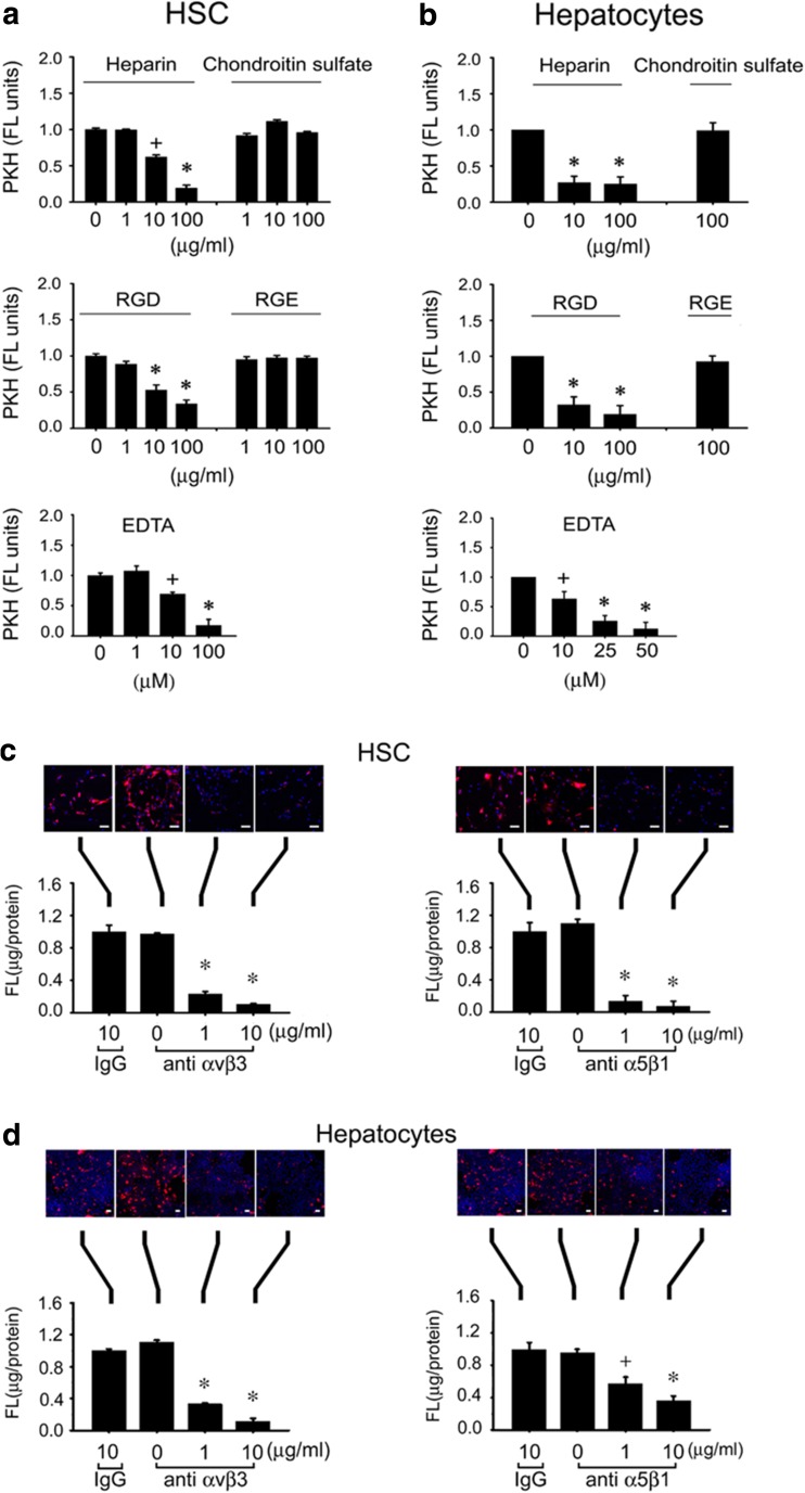
Attenuation of ExoHep binding to hepatocytes or HSC by heparin, RGD tripeptide, EDTA, anti- integrin αvβ3, or anti-integrin α5β1. PKH26-ExoHep were added (8 μg/ml) to (a) P4 HSC or (b) hepatocytes for 24 h in the presence of the indicated concentrations of heparin, chondroitin sulfate, RGD, RGE, or EDTA, prior to measurement of cell-associated fluorescence by spectrophometric quantification of PKH26 in cell lysates (*p < 0.001 vs. “0”, +p < 0.05 vs. “0”, student’s t-test). (c) P4 HSC or (d) hepatocytes were pre-treated for 1 h with 0–10 μg/ml anti-integrin αvβ3, 0–10 μg/ml anti-integrin α5β1, or 10 μg/ml non-immune IgG after which the cells were extensively rinsed and then incubated for 24 h with 8 μg/ml PKH26 ExoHep . Cell-associated fluorescence was determined by confocal microscopy (upper panel) or spectrophotometric quantification of cell lysates (lower panel). Data are from triplicate determinations from 3 or 4 individual experiments. (*p < 0.001 vs. “0”; +p < 0.05 vs. “0”, student’s t-test). Scale bars: 20 μm

