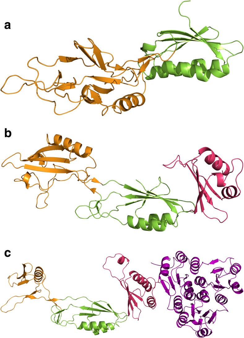
Fig. 4.
Adhesin structures. a LMW SLP: 3CVZ (Fagan et al. 2009). b Cwp2: 5NJL (Bradshaw et al. 2017a). c Cwp8: 5J7Q (Usenik et al. 2017). Cwp2 and Cwp8 assume similar folds with domain 2 rotated approximately 40°. Domain 2 of LMW SLP has significantly longer loop regions and is positioned differently to that of Cwp2 and Cwp8. LMW SLP is covalently bound to HMW SLP so it is likely that domain 3 of LMW SLP is at least somewhat different to that of Cwp2 and Cwp8. Domain colours follow those given in Fig. 3

