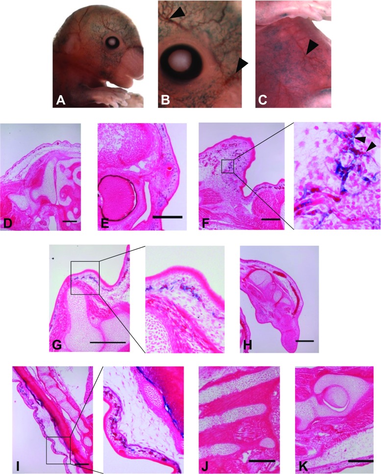
Fig. 2.
-100 kb upstream sequence of CCN2 gene drives transgene activity within microvasculature. Larger blood vessels were not stained (arrowheads b and c) contrasting the punctate staining in the surrounding area. Transgene localisation was concomitant with that of microvasculature and capillary vessel endothelium, with erythrocytes visible within blue branched structure (f, black arrowheads). This pattern was observed globally in a highly superficial manner such as in the frontal portion of the craniofacial region (e and f) and primordial dermis surrounding the fore limb (g) and spine (i). Staining was not observed within other populations of endothelial cells, such as in the growth plate of endochondral tissue. There was an absence of staining in any other tissue type including cartilaginous tissue of the chondrocranium (d and e), wrist (h), intervertebral disc (i), ribs (j) and femur (k). Early osseous tissue within these regions was also negative for transgene expression (Bar = 100 μm)

