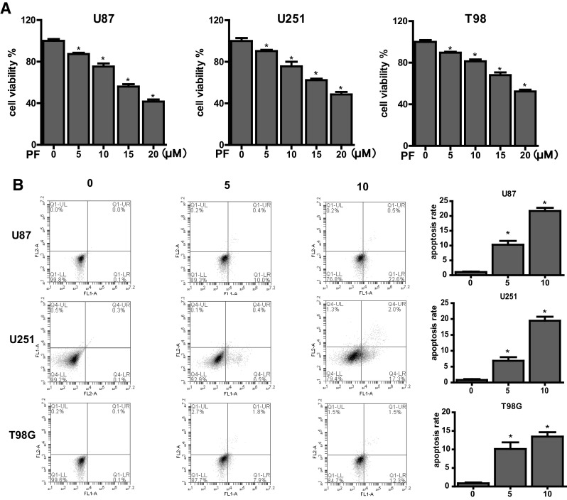
Fig. 1.

Effects of paeoniflorin (PF) on proliferation, apoptosis in U87, U251, T98 cells. a Cells were incubated with the indicated concentrations of PF for 24 h before CCK-8 assay. b Cell apoptosis in glioblastoma cells treated with PF was determined by flow cytometry. Each treatment was replicated at least three times. All tests were performed in triplicate and presented as mean ± standard error. *P < 0.05, compared with control (0 µM)
