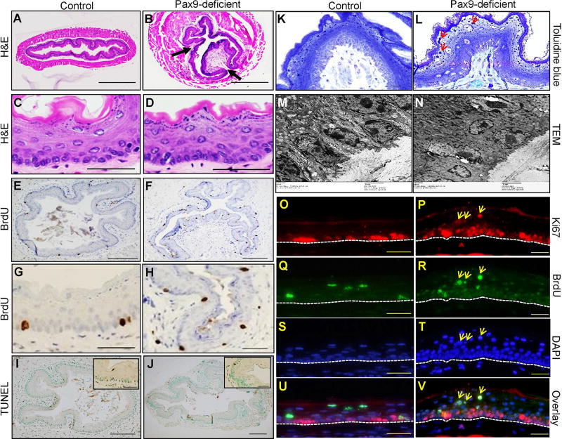
Figure 1. Esophageal phenotypes of Krt5Cre;Pax9loxP/loxP mice.
The papilloma-like structures were formed in the squamous epithelium (indicated by arrows) of Pax9-deficient esophagus (B, D) as compared with control esophagus on H&E-stained sections (A, C). Proliferation was increased in Pax9-deficient mice (F, H) as compared with controls (E, G). Apoptosis was similar in Pax9-deficient esophagus (J) and control esophagus (I), as detected by TUNEL assay. Toluidine blue staining (K, L) and transmission electron microscopy (M, N) showed an increase in keratohyalin granules (red arrows) and disorganization of the basal cell layer in Pax9-deficient esophagus as compared with control esophagus. Double IF showed that Ki67 was exclusively expressed in the basal cells of esophageal epithelium in control esophagus (O, Q, S, U) at the fifth day after BrdU pulse injection. However, in the mutant esophagus, some Ki67 positive cells appeared in the parabasal and superficial layers, and they were also BrdU-positive with the same staining intensity as the BrdU-positive cells in the basal cell layer (P, R, T, V) (yellow arrows). The base membrane of the squamous epithelium was marked with white dotted lines. (A, B, E, F, I, J: Scale bar=200 µm. C, D, G, H, O, P, Q, R, S, T, U, V: Scale bar=50 µm. K, L: Scale bar=20 µm. M, N: Scale bar=2 µm).

