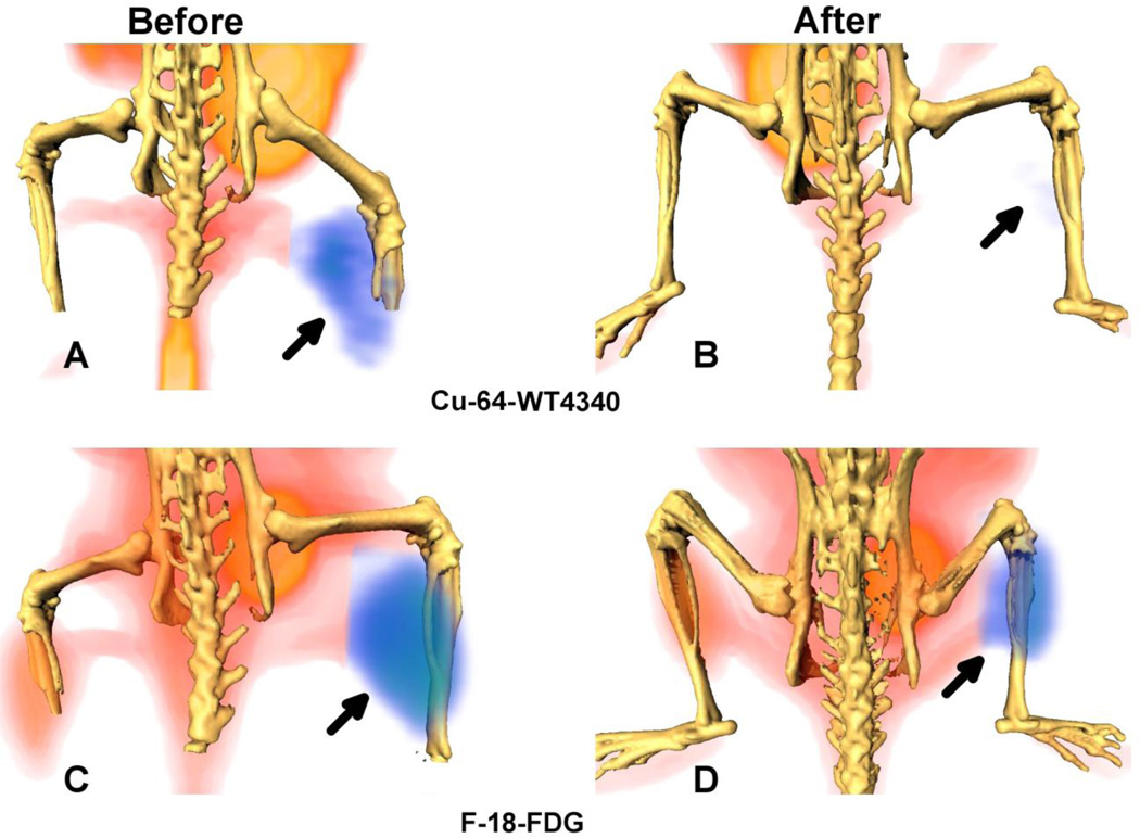
Figure 2.
Surface rendered F-18-FDG and Cu-64-WT4340 images of a mouse bearing BT474 tumor (arrow) in the right thigh. Images in A and C are before treatment and in B and D are after second Dox treatment. Top: Cu-64-WT4340 (A, B) Bottom: F-18-FDG (C, D). A significantly diminished uptake of Cu-64-WT4340 after Dox treatment is visible in (B) as compared to that with the F-18-FDG uptake (D). Note: significant muscle uptake of F-18-FDG in contralateral normal thigh (C).

