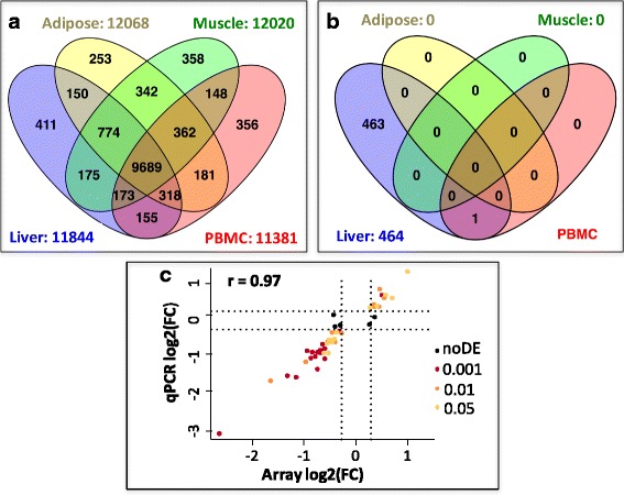
Fig. 2.

Overview of gene expression and differential expression between diets in liver, adipose tissue, muscle and PBMC. a. Number of genes expressed in the 4 tissues. b Number of genes differentially expressed between diets in the 4 tissues. c Pearson correlation between expression fold change in liver (log2(FC)) for 50 genes analyzed by microarray (x-axis) and RT-qPCR (y-axis) in the same experimental design; Significance of the DEG by microarray is indicated by the following color chart: brown dots = p-value ≤0.001, orange dots = p-value ≤0.01, yellow dots = p-value ≤0.05, black dots = node (p.value > 0.05)
