Fig. 2.

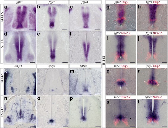
Expression of FGF receptors and FGF target genes at stages of ventral OPC specification in the spinal cord. a-f Expression patterns of mRNA encoding for fgfr1 (a, d), fgfr2 (b, e) and fgfr4 (c, f) on transverse spinal cord sections isolated before OPC specification (E4-E4.5) or at the onset of OPC generation (E5.5-E6). g-h Double detection of Olig2 and fgfr2 (g) or fgfr4 (h) at E5.5-E6. Note expression of both mRNA in Olig2-positive progenitor cells at stages of OPC specification. i-j Double detection of Nkx2.2 and fgfr2 (i) or fgfr4 (j) at E5.5-E6. Note that expression of fgfr2 and fgfr4 mRNAs is restricted to the dorsal-most region of the Nkx2.2 positive domain. k-p Expression patterns of mRNA encoding for mkp3 (k, n), spry1 (l, o) and spry2 (m, p) on transverse spinal cord sections showing that activation of mkp3 in ventral progenitor cells occurs between E4-E4.5 and E5.5-E6 while expression of spry1 and spry2, although detected at low level at E4-E4.5, is reinforced at E5.5-E6. q-r: Double detection of Olig2 and spry1 (q) or spry2 (r). s-t: Double detection of Nkx2.2 and spry1 (s) or spry2 (t) showing expression of both mRNA restricted to the dorsal-most Nkx2.2 positive progenitor cells at stages of OPC specification. Scale bars = 100 μm in a-f and k-p, 50 μm in g-j and k-t
