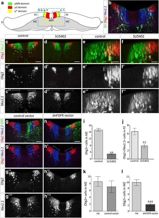
Fig. 4.

Inactivation of FGFRs impairs OPC specification. a Scheme of a transverse section through spinal cord explant cultivated as opened-book showing ventral progenitor domains. The dashed blue panels outline the areas shown in images below. b Immunodetection of O4 (green), Olig2 (red) and Nkx2.2 (blue) on transverse section of spinal cord explant cultivated for 2 days in control condition. Note overlapping of the three markers both in the p* domain and in cells emigrating ventrally in the mantle zone. c-d Immunodetection of Olig2 (red in c and d, c', d') and Nkx2.2 (green in c and d, c'', d'') on transverse sections of spinal cord explants cultivated for 2 days in control condition (c) or in presence of SU5402 (d). Note that while Nkx2.2/Olig2-positive OPCs have already invaded the mantle zone in control explants, very few develop in presence of the inhibitor. Note also reduced dorsal extent of the Nkx2.2-positive domain (brackets) in SU5402 treated explant (d”) compared to control explant (c″). e-f Higher magnification of the progenitor zone showing reduced number of Olig2/Nkx2.2-positive cells in the progenitor zone of explant treated with SU5402 (f, f', f'') compared to control explant (e, e', e''). g-h Immunodetection of Olig2 (red in g, g', h and h', g", h'') and Nkx2.2 (blue in g, h, g' and h', g''', h''') after electroporation of control (green in g) or dnFGFR (green in h) vectors. Note reduction of the dorsal extent of the Nkx2.2 positive domain on the side of explant electroporated with the dnFGFR vector compared to the non electroporated side of the explant or with explant electroporated with the control vector (brackets in g' and h'). Note also that migrating Olig2/Nkx2.2 double-labeled cells are not detected on the side of explant electroporated with the dnFGFR vector. i Quantification of Olig2-positive cells in the mantle zone in control conditions (n = 7) or in presence of the FGFR inhibitor SU5402 (n = 8). j Quantification of Nkx2.2/Olig2-positive cells in the progenitor zone (PZ) of explants cultivated in control conditions (n = 7) or in presence of SU5402 (n = 8). k-l Quantification of Olig2-positive cells migrating in the mantle zone on each side of the explants after electroporation of control (n = 4, k) or dnFGFR (n = 9, l) vectors. Results are presented as mean number of cells ± sem (**p ≤ 0.01; *** p ≤ 0.001). ne: non-electroporated side of explants. PZ = progenitor zone, MZ = mantle zone, MFP = medial floor plate, V = ventral, D = dorsal. Scale bars = 50 μm in b-d, g, h and 25 μm in e, f
