
Fig. 3.
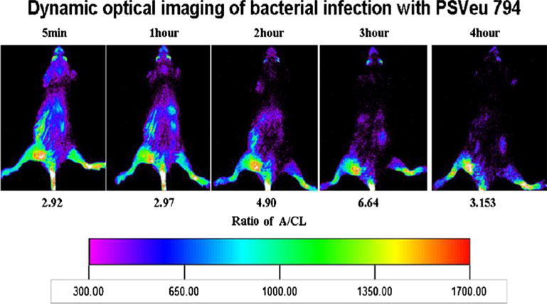
Dynamic optical images of one mouse bearing bacterial infection were acquired for 10 s at 5 min and at 1, 2, and 3 h post-tail vein injection of PSVue®794. Ratios of intensity in abscess to contralateral thigh (A/CL) are given. Declining uptake in liver and kidneys is visible as a function of time after injection.