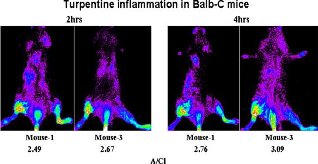
Fig. 4.

Posterior optical images of mice with turpentine-induced inflammation. Ten-second images were acquired at 2 and 4 h post-tail vein administration of 2.7 pmol of PSVue®794. During intravenous injection, a small quantity of PSVue®794 was infiltrated, which is visible. The presence of the probe in the left leg was external contamination (x-ray images are not shown).
