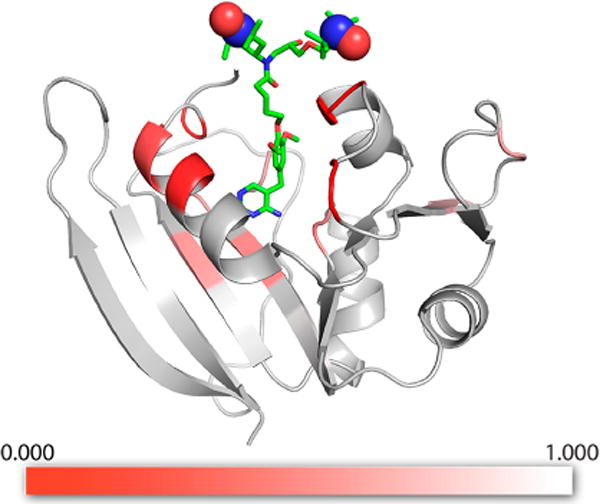
Figure 6.

DHFR model with low temperature SSNMR bleaching information shown as a heat map. The ratio of signal intensity for specifically isotopically enriched DHFR samples with bound TMP-V-T relative to samples with exogenous AMUPol/TMP, observed using DNP-enhanced 13C–13C DARR spectra (ITMPVT/IAMUPol), is indicated by the color scale, red to white. The strongest reduction in signal intensity, or bleaching, is observed for residues proximal to the TMP-V-T binding pocket. The structure of the TMP-V-T linker and TOTAPOL was built into the crystal structure by hand using the Maestro software (Schrödinger Release 2017-3: Maestro, Schrödinger, LLC, New York, 2017), with the paramagnetic N–O groups represented as spheres. The image was produced using the PyMOL Molecular Graphics System, version 1.8.4.2, Schrödinger, LLC.
