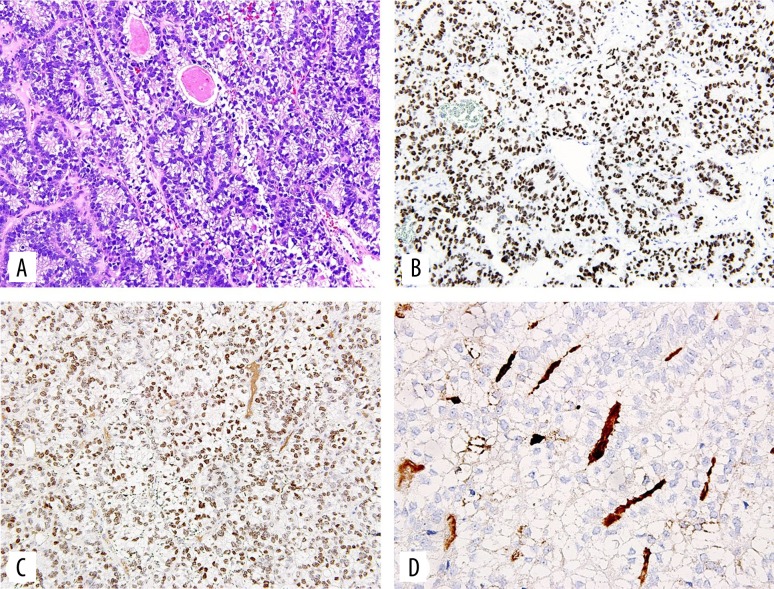
Figure 2.
(A) Light microscopy on hematoxylin and eosin stained slide shows a tumor displaying a glandular, enteric like growth pattern with prominent subnuclear and supranuclear clear vacuoles within the cuboidal or cylindrical cells. (B) Immunohistochemistry with the antibody to SALL4 shows nuclear staining. (C) Immunohistochemistry with the antibody to CDX2 shows nuclear staining. (D) Immunohistochemistry with the antibody to alpha fetoprotein shows focal staining of the intraluminal material in some glands, (160×).

