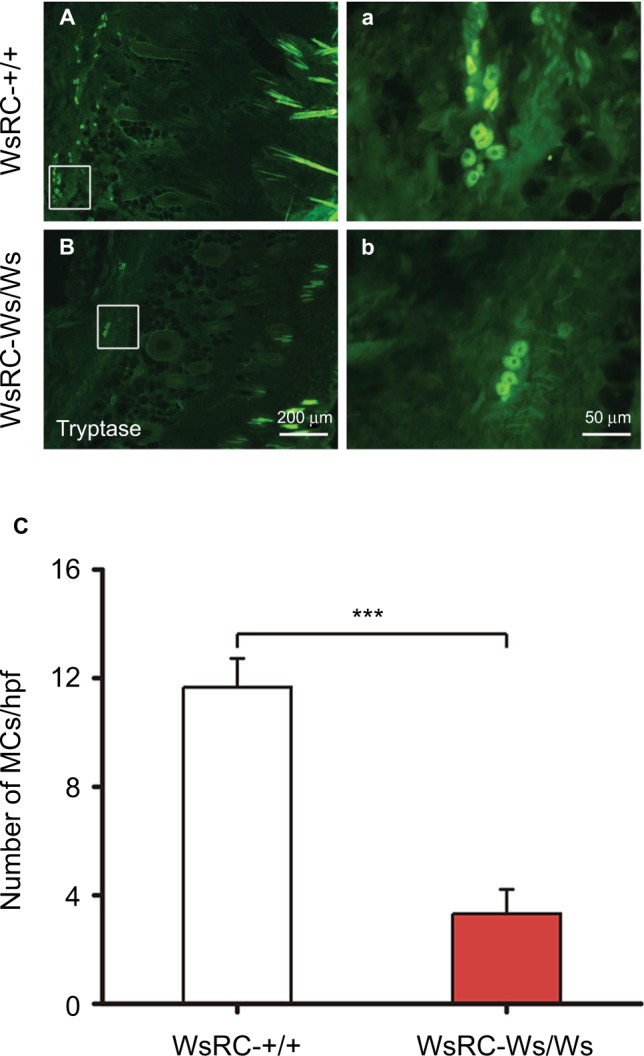
Figure 1.

Immunofluorescence staining for tryptase (MCs) in ST36 skin tissue.
Notes: Representative images of MC staining in WsRC-+/+ (A, a) and WsRC-Ws/Ws (B, b) rats (n=5). (C) MC count in mutant rats was significantly less than that of WT rats. Data were analyzed using independent t-test and expressed as mean ± standard error of the mean. ***P<0.001, vs WsRC-+/+ rats.
Abbreviations: MCs, mast cells; WT, wild-type.
