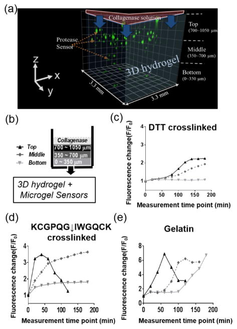
Figure 5.
(a) Three-dimensionally reconstituted image of microgel sensors encapsulated in various hydrogels. A collagenase solution was applied to the top of hydrogel and changes in F/F0 of the microgel sensors were analyzed as a function of space and time. Microgel sensors were categorized according to their location in the z-direction as top (700~1050 μm), middle (350~700 μm) and bottom (0~350 μm). (b,c,d) Spatiotemporal fluorescence change (F/F0) of microgel sensors after an exposure to a 5 mg/ml of collagenase solution on the top of (c) DTT crosslinked PEG hydrogels and (d) KCGPQG↓IWGQCK crosslinked PEG hydrogels and (e) gelatin hydrogels.

