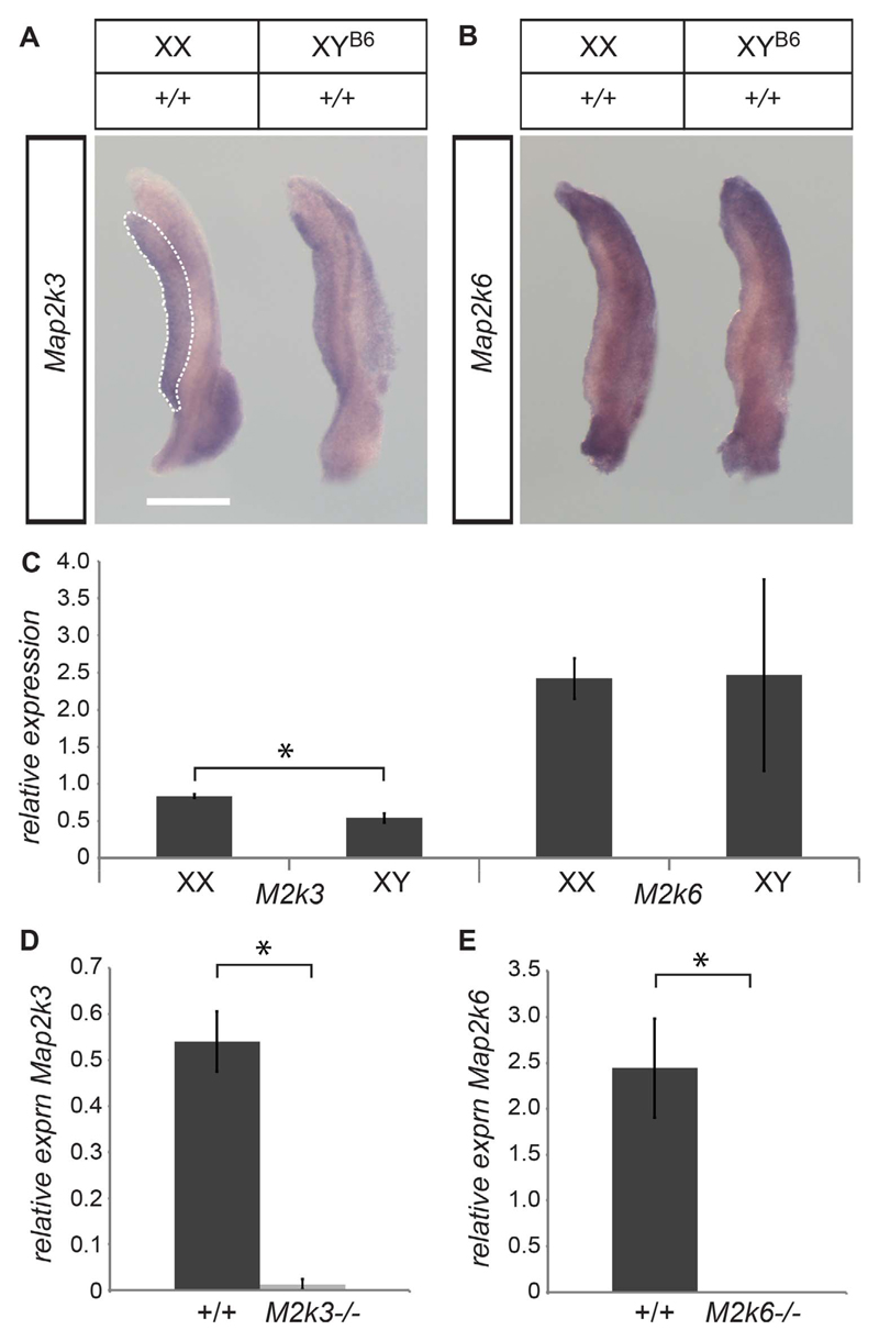
Fig. 1. Map2k3 and Map2k6 are expressed in the developing mouse gonad at the sex-determining stage.
A) Wholemount in situ hybridisation (WMISH) at 11.5 dpc showing Map2k3 expression in the gonad (area within the white dotted lines) and, to a lesser extent, the adjacent mesonephros, with prominent expression in the Wolffian duct. No sexual dimorphism is evident. Bar = 500 μm. B) WMISH at 11.5 dpc showing widespread expression of Map2k6 in the developing gonad and mesonephros, with stronger signal detected in the mesonephros. C) Comparison of expression of Map2k6 and Map2k3 in XY and XX wild-type gonads. Map2k3 expression in XX gonads is higher than XY gonads by a small, but significant, amount. Error bars throughout show standard error of the mean; *P < 0.05. D) Quantitative RT-PCR was performed using primers that amplify exonic sequences that are deleted in the Map2k3 null allele. Expression detectable in wild-type gonads is all but absent in gonads from MAP2K3-deficient embryos, validating the specificity of the expression measurement. E) Quantitative RT-PCR was performed using primers that amplify exonic sequences that are deleted in the Map2k6 null allele. Expression detectable in wild-type gonads is absent in gonads from MAP2K6-deficient embryos. RNA expression levels were normalized to those of Hprt1 using the ΔΔCt method. Error bars show standard error of the mean.

