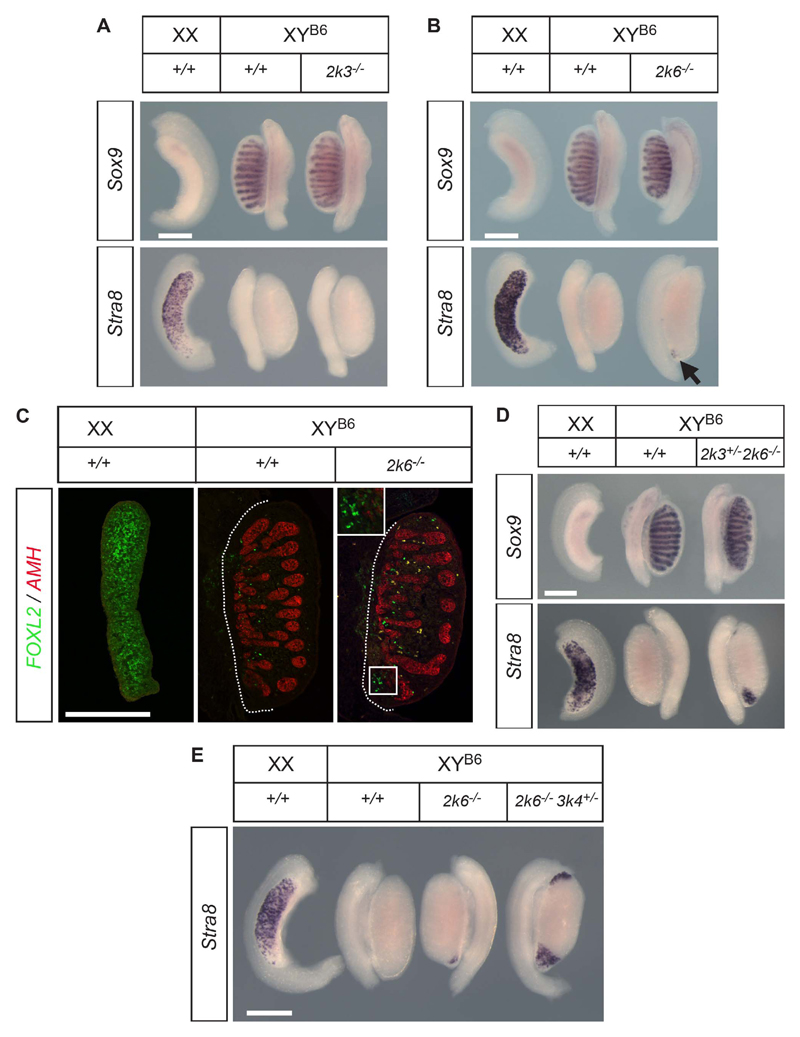
Fig. 2. Characterization of gonadal phenotypes at 14.5 dpc in B6 embryos lacking MAP2K3 and MAP2K6.
A) WMISH of Sox9 and Stra8 in gonads from MAP2K3-deficient embryos reveals no overt abnormalities. Bar = 500 μm. B) WMISH of Sox9 and Stra8 in gonads from MAP2K6-deficient embryos reveals a small but consistent patch of Stra8 expression (arrow) at one pole of the mutant gonad. Bar = 500 μm. C) Immunostaining of gonadal tissue sections comparing MAP2K6-deficient B6 XY embryos and wild-type controls with the Sertoli cell marker AMH (red) and granulosa cell marker FOXL2 (green) reveals a small cluster of FOXL2-positive cells at the caudal pole of the mutant (2k6−/−) gonad (white box) exhibiting nuclear staining (see higher magnification image in inset). A large number of fluorescent cells in the interstitium of the mutant gonad are blood cells. The occasional interstitial cell can also be seen exhibiting nuclear staining, but these rare cells are also observed in wild-type control gonads and are of unknown significance. The dotted line indicates the border between the gonad (right) and mesonephros (left). Bar = 500 μm. D) Compound mutant embryos (Map2k3+/−, Map2k6−/−) have gonads exhibiting a larger area of Stra8 expression at one pole, indicating some redundancy between MAP2K3 and MAP2K6 function during testis determination. Bar = 500 μm. E) WMISH reveals more extensive regions of Stra8 expression at the gonadal poles in compound mutants (Map2k6−/−, Map3k4+/−) when compared to littermates only lacking Map2k6 (Map2k6−/−). Bar = 500 μm.

