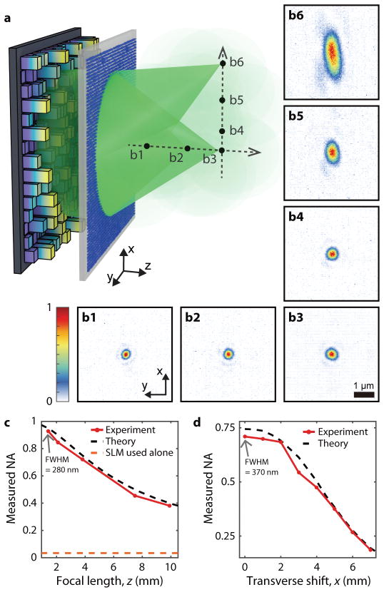
Figure 3. Experimental demonstration of diffraction-limited focusing over an extended volume.
(a) Schematic of optical focusing assisted by the disordered metasurface. The incident light is polarized along the x direction. (b1–6) Measured 2D intensity profiles for the foci reconstructed at the positions indicated in (a). b1–b3 are the foci along the optical axis at z = 1.4, 2.1, and 3.8, mm, respectively, corresponding to NAs of 0.95, 0.9, and 0.75. b3–b6 are the foci at x = 0, 1, 4, and 7 mm scanned on the fixed focal plane of z = 3.8 mm. Scale bar: 1 μm. (c) Measured NA (along x-axis) of the foci created along the optical axis (red solid line) compared with theoretical values (black dotted line). When the SLM is used alone, the maximum accessible NA is 0.033 (orange dotted line), based on the Nyquist-Shannon sampling theorem. (d) Measured NA (along x-axis) of the foci created along x axis at z = 3.8 mm (red solid line) compared with theoretical values (black dotted line). The number of resolvable focusing points within the 14-mm diameter FOV was estimated to be 4.3×10⁸.

