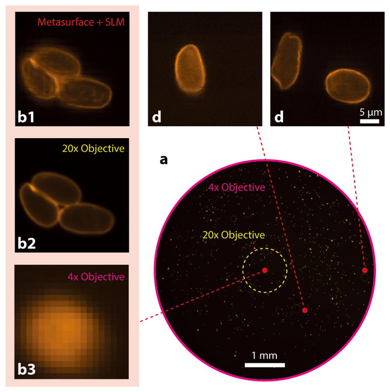
Figure 4. Demonstration of disordered metasurface assisted microscope for high resolution wide-FOV fluorescence imaging of giardia lamblia cysts.

(a) Low resolution bright field image captured by a conventional fluorescence microscope with a 4× objective lens (NA = 0.1). Scale bar: 1 mm. (b1–3) Fluorescence images captured at the center of the FOV. (b1) Scanned image obtained with a disordered metasurface lens. (b2) Ground truth fluorescence image captured with a 20× objective lens (NA = 0.5). (b3) Magnified low-resolution fluorescence image captured with the 4× objective. (c, d) Images obtained with the disorder metasurface-assisted microscope at (x, y) = (1, 1) and (2.5, 0) mm, respectively. This demonstrates that we can indeed use the system for high resolution and wide FOV imaging.
