Figure 1.
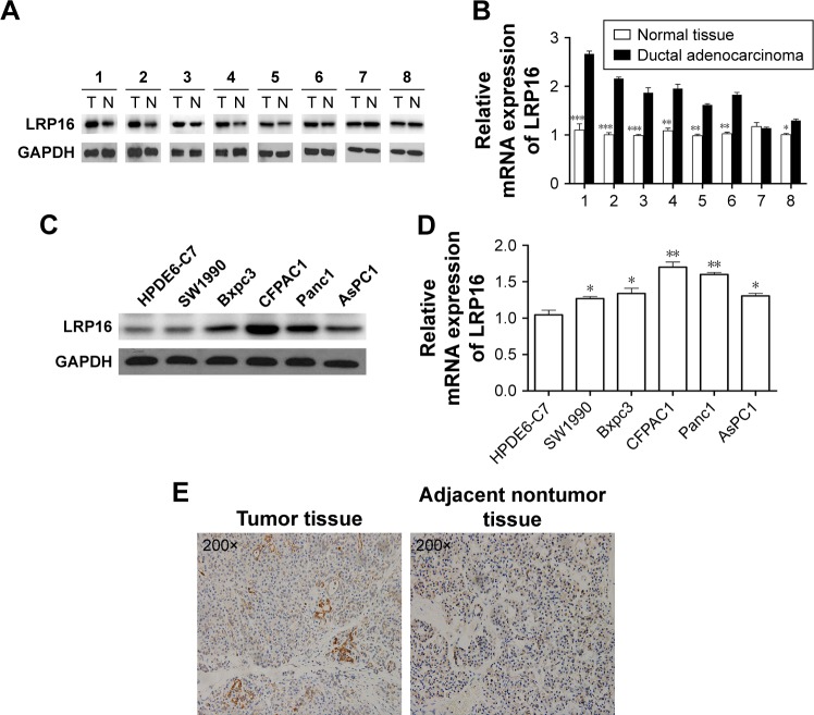
LRP16 expression in pancreatic cancer cell lines and surgical specimens.
Notes: (A) The expressions of LRP16 in eight pairs of pancreatic cancer tissues and normal pancreatic tissues were evaluated by Western blotting (N = normal liver tissues, T = tumor tissues). (B) The mRNA levels of LRP16 expression in eight pairs of pancreatic cancer tissues and normal pancreatic tissues were detected by qPCR. Data shown represent mean ± SD for triplicate samples from a representative experiment. *P<0.05; **P<0.01; ***P<0.001. (C and D) Normal pancreatic cell line HPDE6-C7 and five pancreatic cancer cell lines were subjected to Western blotting and qPCR to determine the intracellular LRP16 expression, and GAPDH was used as loading control. (E) Immunohistochemistry for LRP16 expression in pancreatic cancer samples and nontumor samples (200×).

