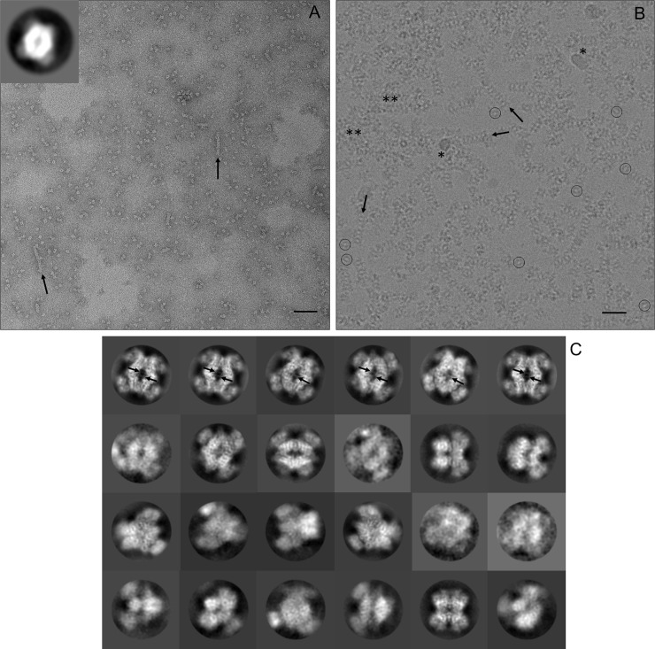
Fig 2. Raw micrographs and 2D class averages.
(A) A section of a negative stained grid imaged with an actual magnification of 50,607x is shown. The inset in the top left corner shows the 2D class average corresponding to over 50% of the observed particles. The diamond-like shape of the PRMT5 core is well-resolved, but the MEP50 is poorly resolved. The bottom right scale bar is 90 nm. (B) A section of PRMT5:MEP50 particles embedded in vitreous ice is shown. The image is a motion corrected summation of 40 individual movie frames at an actual magnification of 50,000x. A low contrast, central ‘donut hole’ is visible in individual particles (circled). Particles of similar orientation to the (A) inset appear like ‘headless turtles’. Ice and/or ethane contamination and aggregated protein are indicated by * and **, respectively. The bottom right scale bar is 30 nm. (C) Representative 2D class averages calculated from cryo-images are shown with inverted contrast. The maximal PRMT5:MEP50 particle dimension is approximately 15 nm. Black arrows highlight small filamentous aggregates in (A) and (B), and pore loop density for residues 488–493 in (C).

