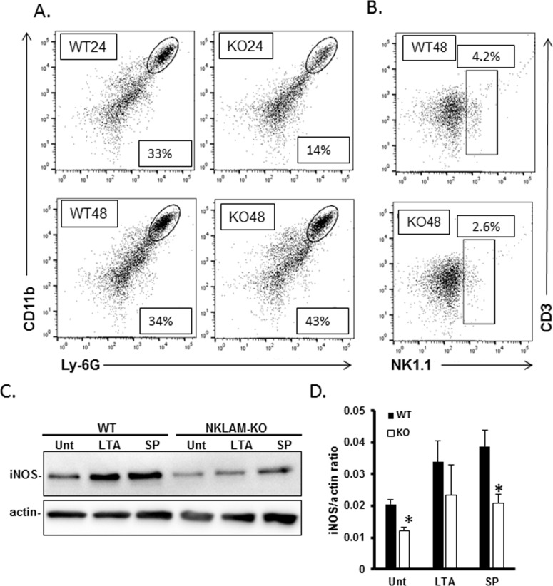
Fig 3. Neutrophil and NK cell lung infiltration and bone marrow-derived macrophage iNOS expression.
Cells were isolated from infected lungs at 24 and 48h post infection and stained for CD45, CD3, NK1.1, CD11b and Ly-6G. CD45+ cells were gated and the percentage of CD11b+/Ly-6G+ (A) or CD3-/NK1.1+ (B) cells within the CD45+ population was determined. A representative histogram is shown for each group (n = 3–4 mice per group). (C) BMDM were treated with 100 μg/mL lipoteichoic acid (LTA) or formalin-fixed S. pneumoniae (SP) at an MOI of 10 for 18 hr at 37°C and protein lysates were immunoblotted for iNOS protein. Beta actin was used as a loading control. Immunoblots represent 1 of 3 identical experiments, (D) Graphical representation of C. n = 3; *p < 0.05.

