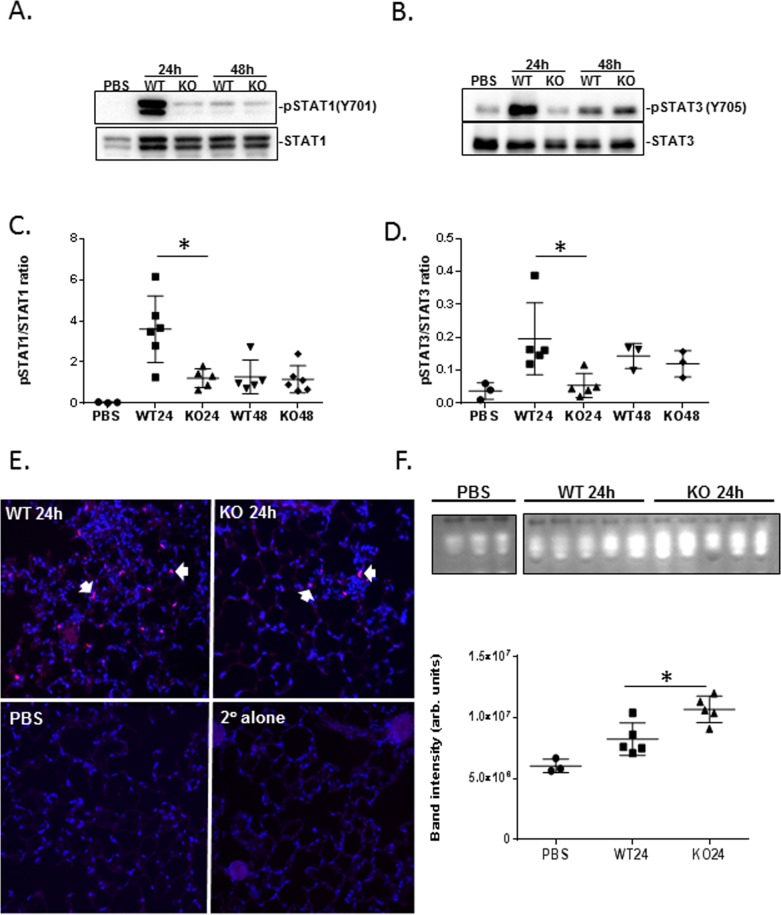
Fig 6. S. pneumoniae-induced phosphorylation of STAT1 and STAT3 is diminished in NKLAM-KO mice.
Lung homogenates from S. pneumoniae-infected WT and NKLAM-KO mice were immunoblotted for (A) STAT1 and pSTAT1 (Tyr701) and (B) STAT3 and pSTAT3 (Tyr705). Each lane represents an individual mouse and is representative of the overall experiments. Immunoblots were used to calculate the ratio of pSTAT1 (Tyr701)/STAT1 (C) and pSTAT3 (Tyr705)/STAT3 (D). Data represent the mean ± SD of 1 of 3 representative experiments (n = 3–5 mice per group per experiment); * p ≤ 0.05. (E) Sections from paraffin-embedded lungs from S. pneumoniae-infected (24h) WT and NKLAM-KO mice were stained for pSTAT1 (Tyr701) (red) and DAPI (blue). Colocalization is depicted in pink (arrows). (n = 2–3 mice per group). (F) Aliquots (50 μg) of lung homogenate from PBS control mice and S. pneumoniae-infected (24h) WT and NKLAM-KO mice were separated by native gel electrophoresis then incubated with 4-methylumbelliferyl phosphate to visualize phosphatase activity. Each lane represents an individual mouse. Graph represents the mean band intensity ± SD. Results are representative of two experiments with 3–5 mice per group per experiment; * p ≤ 0.05.

