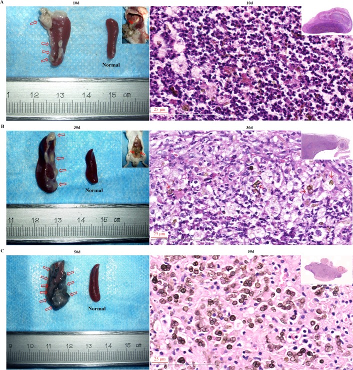
Fig 3. Recalcitrant infection of BALB/c mice was induced by intraperitoneal injection of F. pedrosoi hyphae with the formation of sclerotic cells in the nidus.
(A-C) Spleens respectively from BALB/c mice at 10 days (A), 30 days (B) and 50 days (C) post-inoculation as well as the normal control (left column). The infectious foci in the spleens and abdomen were indicated by red arrows. Scale bar = 1 mm. (A-C) HE staining (×400) for the infected spleens at 10 days (A), 30 days (B) and 50 days (C) post-inoculation (right column).

