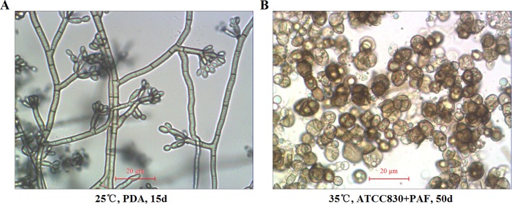
Fig 4. In vitro induced transformation of saprophytic F. pedrosoi into sclerotic cells.
Optical microscope was used to characterize the morphology of saprophytic F. pedrosoi growing on PDA (A) and in-vitro transformed sclerotic cells cultured in ATCC830 medium plus PAF for 50 days (×400) (B). Scale bar = 20 μm (A and B).

