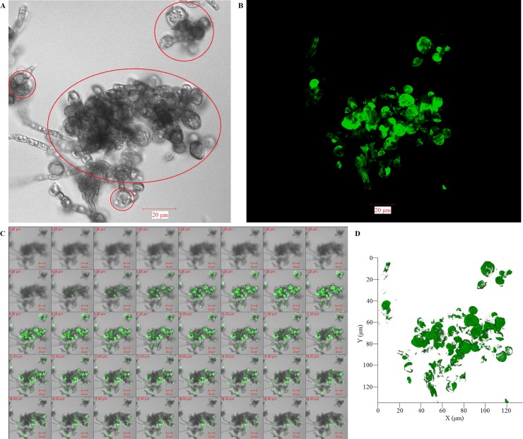
Fig 11. Three-dimensional conformation of chitin distribution re-constructs the figuration of in-vitro transformed sclerotic cells.
(A-D) Three-dimensional distribution of chitin on the surface of F. pedrosoi cultured in ATCC 830 medium with 10−6 M PAF for 40 days were analyzed by FITC-conjugated WGA using confocal tomoscanning. (A) F. pedrosoi in the bright field. In-vitro transformed sclerotic cells with cross-septation were indicated by red circles. (B) Three-dimensional conformation of chitin distribution on F. pedrosoi was represented by FITC-conjugated WGA in the fluorescence field. (C) The binding of FITC-conjugated WGA on the surface of F. pedrosoi was analyzed by confocal tomoscanning. Each cross-section thickness was set as 0.5μm. Scale bar = 20 μm. (D) Three-dimensional conformation of chitin distribution represented by FITC-conjugated WGA was reconstructed according to confocal tomoscanning.

