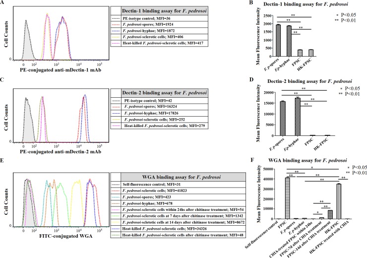
Fig 12. Chitin accumulation on the surface of F. pedrosoi-sclerotic cells compensatorily increased after chitinase treatment in a time-dependent manner.
(A-F) Abbreviations: F. pedrosoi-spore (F. p-spore); F. pedrosoi-hyphae (F. p-hyphae); In-vitro transformed F. pedrosoi-sclerotic cells in ATCC 830 medium with 10−6 M PAF for 50 days (FPSC); Heat-killed FPSC (HK-FPSC); Chitinase (CHIA). (A and B) The binding capacity of murine Dectin-1 to F. p-spores, F. p-hyphae, FPSC or HK-FPSC was respectively detected by PE-conjugated anti-murine Dectin-1 mAb using flow cytometry, and was represented as mean fluorescence intensity (MFI). (C and D) The binding capacity of murine Dectin-2 to F. p-spores, F. p-hyphae, FPSC or HK-FPSC was respectively detected by PE-conjugated anti-murine Dectin-2 mAb using flow cytometry, and was represented as mean fluorescence intensity (MFI). (A-D) The fungal cells incubated only with PE-conjugated isotypes were set as blank control. (E and F) The binding capacity of FITC-conjugated WGA to F. p-spore, F. p-hyphae, FPSC or HK-FPSC before and after chitinase treatment at indicated time points was detected by flow cytometry, and was represented as mean fluorescence intensity (MFI). (B, D and F) Data represent the mean±SEM from three individual experiments performed in triplicates, and statistical analysis was performed using Univariate ANOVA and LSD-t test. Significant: * P<0.05; Highly Significant: ** P<0.01.

