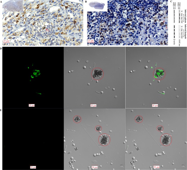
Fig 13. Exclusive accumulation of chitin was also detected on the surface of in-vivo transformed F. pedrosoi-sclerotic cells which were isolated from the nidus with chitinase expression.
(A and B) Expression of acidic mammalian chitinase (AMCase) in the footpads of nu/nu-BALB/c mice at 80 days (A, n = 3), or in the spleens of BALB/c mice at 50 days (B, n = 3) after inoculation with F. pedrosoi was detected by rabbit anti-AMCase polyAb using immunohistochemistry. In vivo-transformed sclerotic cells in the nidus at 50 days after inoculation with F. pedrosoi-hyphae were indicated by red arrows. (C) AMCase expression in the infected footpads of nu/nu-BALB/c mice or in the infected spleens of BALB/c mice was detected by rabbit anti-AMCase polyAb using western blotting at indicated days after inoculation with F. pedrosoi-hyphae (n = 5 per subgroup of BALB/c and nu/nu- BALB/c mice). The nu/nu-BALB/c or BALB/c mice subcutaneously or intraperitoneally inoculated with NS were set as normal controls (n = 15). β-actin in the infected footpads or spleens was set as loading control, and markers were shown. (D) Confocal microscope was introduced to detect the binding of FITC-conjugated WGA to F. pedrosoi isolated from the infected spleens of BALB/c mice at 50 days post-inoculation. (E) F. pedrosoi isolated from infected spleens of BALB/c mice at 50 days post-inoculation with chitinase pretreatment was set as control. In-vivo transformed sclerotic cells were indicated by red circles. Left column: fluorescence field; Middle column: bright field; Right column: merged images. Scale bar = 20 μm.

