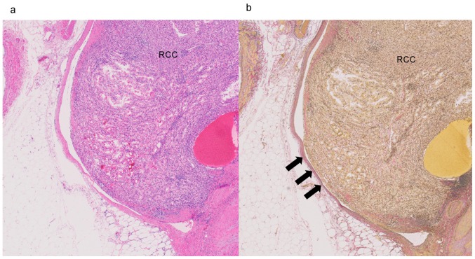
Figure 2.
Photomicrograph of microvascular invasion. (a) Renal cell carcinoma (RCC) is seen protruding into the vessel (hematoxylin and eosin staining; magnification, ×20). (b) The tumor cells broke through the vessel collagen wall and invaded into the vessel (arrows). Elastica Van Gieson staining; magnification, ×20.

