
Fig. 1.
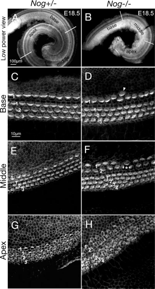
Increased inner and outer hair cells in Nog−/− cochlea. A–H: Phalloidin-labeled Nog+/− (A,C,E,G) and Nog−/− (B,D,F,H) cochleae at embryonic day (E) 18.5. A,B: Nog+/− cochlea shows one and three-fourth turns (A), whereas Nog−/− cochlea shows only one turn (B). Representative images from the base, middle, and apical regions are shown for Nog+/− (C,E,G) and Nog−/− cochleae (D,F,H). C,E,G: A Nog+/− cochlea showing the typical one row of inner hair cells (IHC; 1) and three rows of outer hair cells (OHC; 1,2,3), which are less organized at the apex. D,F,H: Nog−/− cochlea showing occasional extra IHC at the base (D) and two rows of IHC (1,2) and four rows of OHC (1,2,3,4) in the middle and apical turns (F,H). Asterisk in F indicates damaged hair cells. Scale bar in A applies to B. Scale bar in C applies to D–H.