
Fig. 3.
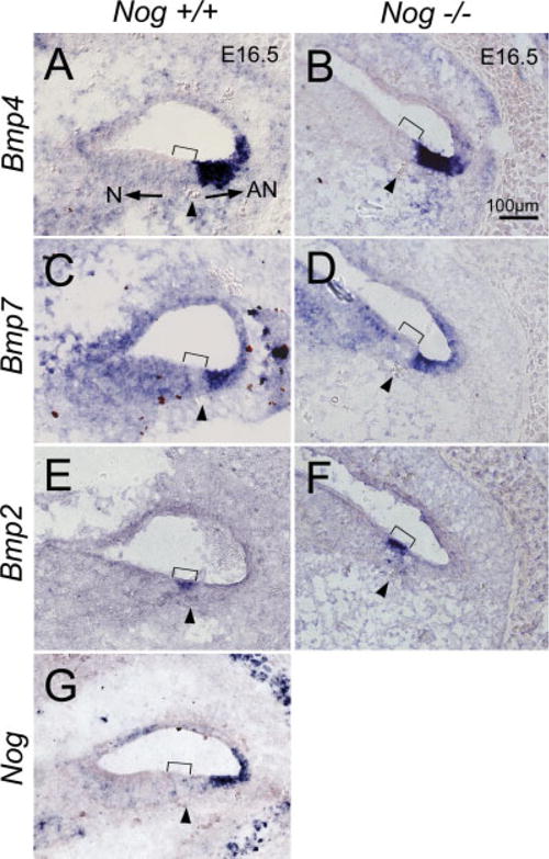
Expression patterns of Bmps and Nog in wild-type and Nog−/− cochleae at embryonic day (E) 16.5. A: Bmp4 is strongly expressed in the abneural region of wild-type cochlea. C: Bmp7 expression is broad but its strongest expression domain overlaps with the Bmp4-positive region. E: Bmp2 is expressed in the sensory region of the cochlea, not overlapping with the Bmp4-positive region. G: The expression domain of Nog partially overlaps with Bmp4 expression. B,D,F: There is no noticeable change in expression patterns of Bmp4, Bmp7, and Bmp2 in Nog−/− cochleae, respectively. Arrowhead indicates the spiral blood vessel beneath the prosensory domain (bracket). Arrows in A point toward neural (N) and abneural (AN) sides of the organ of Corti. Scale bar in B applies to A–G.