
Fig. 5.
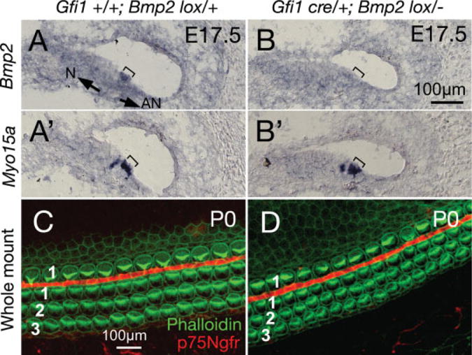
Targeted deletion of Bmp2 using Gfi1cre/+ mice. A–A′: Serial sections of a Gfi1+/+;Bmp2lox/+ cochlea at embryonic day (E) 17.5, showing Bmp2 expression overlapping with Myo15a in outer hair cell (OHC; bracket). B–B′: Serial sections of a Gfi1cre/+;Bmp2lox/− cochlea at E17.5 showing the absence of Bmp2 expression in OHC (compare bracketed regions). C,D: Whole-mounts of Gfi1+/+;Bmp2lox/+ (C) and Gfi1cre/+;Bmp2lox/− (D) cochleae at postnatal day (P) 0, showing the three rows of OHC and one row of inner hair cell (IHC; phalloidin labeling in green), separated by pillar cells (p75Ngfr staining in red). No difference in hair cell formation and organization are detected in Gfi1cre/+;Bmp2lox/− cochlea. Scale bar in B applies to A–A′,B′, and C applies to D. Arrows in A point toward neural (N) and abneural (AN) sides of the organ of Corti.