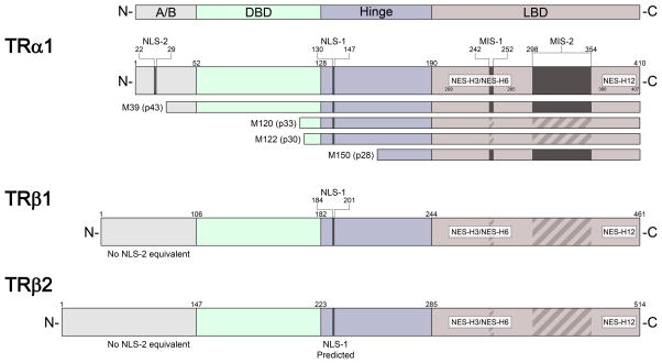
Figure 1.
Thyroid hormone receptor (TR) major subtypes and localization signals. The structural diagram (not to scale) of TRα1, TRβ1, and TRβ2 shows nuclear localization signal (NLS), nuclear export signal (NES), and mitochondrial import sequence (MIS) motifs, where known (solid bar) or predicted (striped bar) based on sequence homology. The positions of localization signals are indicated in relation to the respective individual domains of TR: N-terminal A/B domain (A/B), DNA-binding domain (DBD), hinge domain, and ligand-binding domain (LBD). The TRα1 mRNA encodes several forms of truncated TR by translation initiation from internal AUG sites encoding methionine (M); amino acid residue numbers correspond to the position in full-length TRα1.

