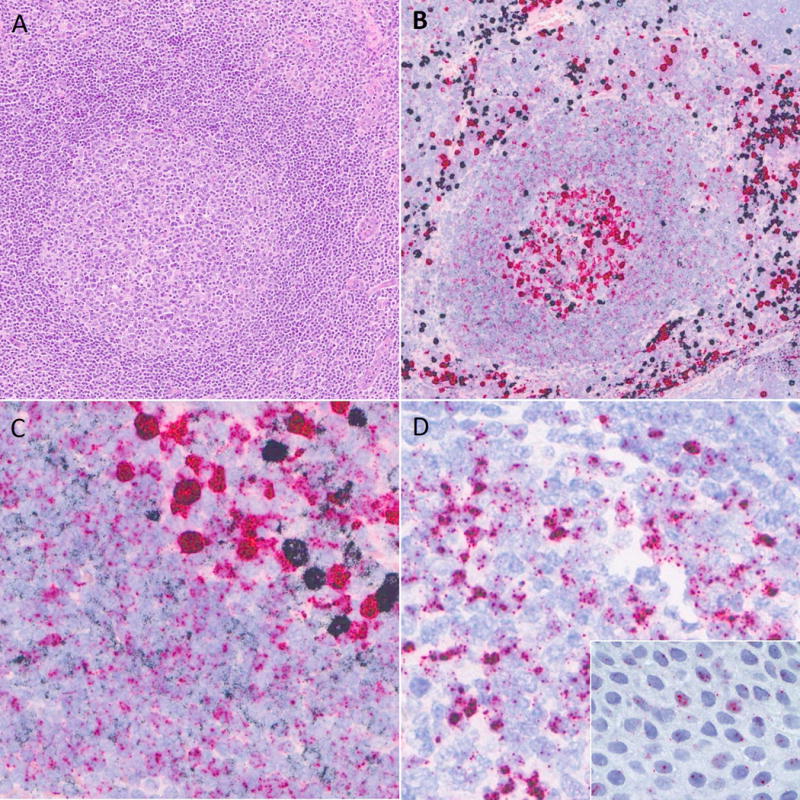
Figure 2.

RNA in situ hybridization staining in normal lymphoid tissues. A) Reactive germinal center in routine H&E. B) Kappa/lambda RNA in situ hybridization shows strongly positive kappa (black) and lambda (red) plasma cells at low power. C) At higher power, polytypic expression is seen in mantle zone lymphocytes (lower left) and germinal center lymphocytes (upper right). D) IGLL5 signal contains nuclear dots and, especially within germinal center lymphocytes, cytoplasmic staining. Inset, IGLL5 nuclear staining in tonsil epithelium.
