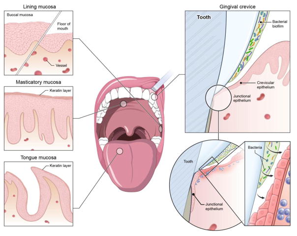
Figure 1. The oral and gingival barrier.
The oral mucosa is lined by stratified squamous epithelia of varying thickness and level of keratinization. Most the oral mucosa is covered by lining, non-keratinized epithelium (the floor of mouth being particularly thin and vascular). The areas related to mastication (hard palate, outer surface of gingiva) are partially keratinized epithelia and the tongue is covered with a specialized epithelium that incorporates the taste buds. The gingival crevice is a particularly open and vulnerable site. It is lined by the sulcular epithelium that is non-keratinized and becomes progressively thinner transitioning to the junctional epithelium, which connects to the tooth surface and is constantly exposed to the microbial biofilm and experiences trauma, leading to constant immune activation.

